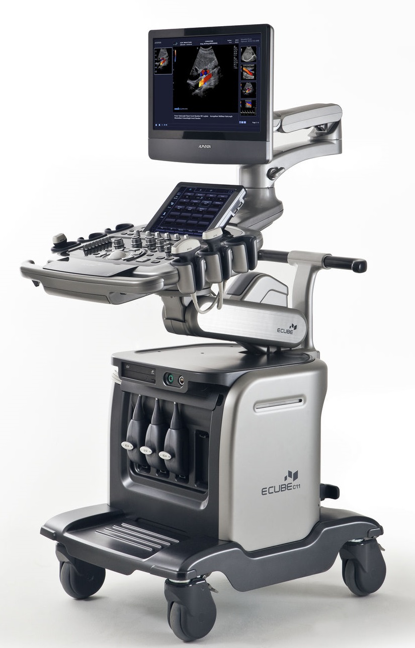
Powerful, Friendly and Compact Ultrasound System
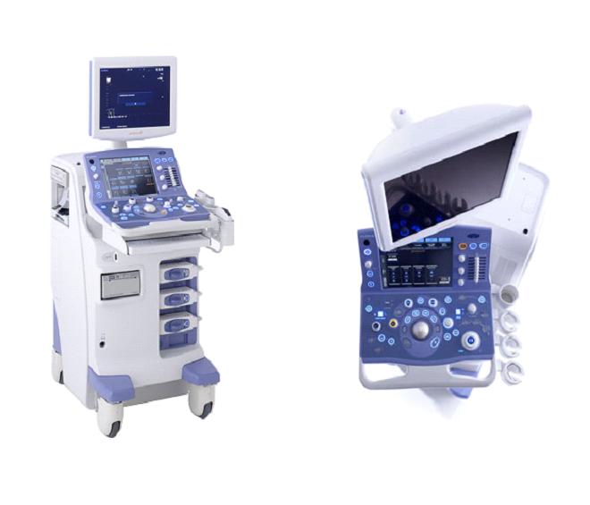
ProSound α7 is a diagnostic ultrasound system realizing efficient examination through high-definition images, ease of use and flexible data management.
It inherits the proven technologies and functions of Aloka's high-end product, yet offers outstanding mobility thanks to being the smallest size in its class. The system is easily transported from the examination room to the ICU, operating theater or patient’s bedside to deliver high performance throughout the hospital.
To reduce the burden on the examiner, the universal design ensures unparalleled ease of use. Improved efficiency of examination also reduces the burden on the patient. And the system is environment-friendly, being made of ecological materials and consuming little power.
Powerful
• Broadband Harmonics was realized, enabling both high spatial resolution and enhanced sensitivity in Harmonic Echo imaging.
• Enhanced color sensitivity
• Directional eFLOW depicts narrow to wide blood vessels precisely without overlapping tissue image
• Real-time 3D with Automated Volume Measurement function
• eTRACKING (Echo Tracking) contributes to preventive medicine by detecting early stage of atherosclerosis
• High-performance cardiac analysis package
Friendly
• User friendly ergonomic design
• Height adjustable and swivel operation panel and LCD monitor
• Patient friendly short examination time
• Environment friendly
• Low power consumption and ecology-conscious materials
Compact
• Lightest in weight in this class
• Small footprint
• High mobility
Scanning Method
• Electronic Convex Sector
• Electronic Linear
• Electronic Phased Array Sector
Beamformer
Transmission
Semi-CPWG (Compound Pulse Wave Generator)
Reception
Multi processing high-speed digital beam former
12-bit A/D converter (4096 gray levels)
Sampling rate: Equivalent to 320 MHz Delay precision: 1/64λ at minimum in both transmission and reception
Transmission/Reception of Harmonics
Broadband Harmonics by Quasi Compound Impulse Waveform Transmission (Probe dependent)
Focusing
Lateral direction
Transmission: Multi-stage transmission focus of up to 4 stages out of 8 stages
Reception: PixelFocusTM
Slice direction
Mompound Dual Focusing is possible (when a Compound Array probe is used)
Beam signal processing
Dynamic apodization
Frame rate
* Max. 700 frames/s or more*
* Depends on probe and parameter settings
Image Display Modes *1
• B: gray-scale imaging
• M
• D: Spectral Doppler (PW, HPRF PW, and CW)
• Dual B
• Quad B
• B and M
• B (Flow)
• B (Power)
• B (eFlow)
• Dual B (Flow)
• Quad B (Flow)
• Dual B (PowerFlow)
• Quad B (PowerFlow)
• Dual B (eFlow)
• Quad B (eFlow)
• M (Flow)
• M (PowerFlow)
• M (eFlow)
• B (Flow) and M (Flow)
• B (Flow) and M (PowerFlow)
• B (Flow) and M (eFlow)
• B (Flow) and D
• B (Flow) and D simultaneous real-time display (Triplex mode)
• B and B (Flow) simultaneous real-time display
(DDD: Dual Dynamic Display)
• B (PowerFlow) and D
• B (PowerFlow) and D simultaneous real-time display (Triplex mode)
• B and B (PowerFlow) simultaneous real-time display
(DDD)
• B (eFlow) and D
• B (eFlow) and D simultaneous real-time display (Triplex mode)
• B and B (eFlow) simultaneous real-time display
(DDD: Dual Dynamic Display)
• B (Flow), M (Flow), and D
• B (PowerFlow), M (PowerFlow), and D
• B (eFlow), M (eFlow), and D
• CHE Intermittent trigger mode *2
• Dual Dynamic Monitor*2 (Fundamental image/CHE image, side by side display)
• TDI (Tissue Doppler Imaging)
• Real-time 3D (4D) mode*3
• Trapezoidal scan
Request function: In multi-mode display, it is possible to select one mode for full screen display.
*1 Probe dependent. *2 Option: CHM-ALPHA7 *3 Option: EU-9121
B-mode
• Display Gray Scale: 256 levels
• Scanning area:
100% to 25%, continuously variable
• Zoom
Write zoom (magnification of real-time image):
Max. 6 times (probe dependent)
Read zoom (magnification of frozen image):
Max. 16 times
• Depth range selections:
0.5/0.75/1.0/1.5/2.0/2.5/3.0/3.5/4.0/5.0, and 1 cm-step increment from 5.0 to 30 cm (probe dependent)
• Longitudinal and lateral inversion
• Rotation by 90 degrees (probe dependent)
• Frame rate (Line density): 3 selections (-1, 0, +1) • Gain*1: 10 to 90 dB
• STC (sensitivity time control) — gain versus depth curve control: 8 slide controls
• Angle gain:
Gain versus angle curve control: 8 sectors (probe dependent)
• Contrast*1: 23 steps (Dynamic range: 36-96 dB) (It is possible to select the indication between dynamic range in dB and number of steps.)
• AGC—Suppression of brightness saturation and Edge Enhancement: 16 steps
• Relief: 4 steps
• FTC: On/Off
• Frame correlation: 16 steps (Auto/Manual)
• Smoothing: 16 steps (1-16) • Post Processing
Echo enhance curve: 5 kinds
Rejection: 64 steps
• View gamma: Off + 4 kinds
• Edge optimizer: 8 steps
• Image optimizer: Available
• STC curve memory: Available
*1 Gain and contrast can be changed after freezing
• Spatial compound scan*2 (possible by selected linear and convex sector probes) Compound scan angle: 5-20 degrees (probe dependent)
*2 SOP-ALPHA7-22 is necessary.
• Trapezoidal scan (possible by selected linear probes)
• Adaptive Image Processing (AIP)*3 *3 SOP-ALPHA7-24 is necessary.
M-mode:
• Sweep method: Moving bar
• Sweep speed*1:
• 200, 150, 100, 66.7, 50, 33.3, 25 mm/sec.
• Gain*1: B-gain ±30 dB
• Contrast*1: 23 steps (Indication can be selected between dynamic range and number of steps.)
• AGC—Suppression of brightness saturation: 16 steps (including relief processing)
• Relief: 4 steps
• FTC: On/Off
• FAM*2 (Free Angular M-mode) Up to 3 M-mode cursors can be set omni-directionally on real-time at any position on a B-mode image.
It is possible to reconstruct M-mode images from line-data B-mode images stored in the HDD. *1 Gain, contrast and sweep speed are changeable after freezing
*2 Optional: SOP-ALPHA7-5
Spectral Doppler:
• Display: Power spectrum
• Real-time Doppler Auto Trace: Possible*
* Option: SOP-ALPHA7-3
• Doppler methods:
• PW (Pulsed Wave) Doppler
• HPRF (High Pulse Repetition Frequency) PW Doppler
• CW (Continuous Wave) Doppler*
* Option: UCW-ALPHA7
• Reference frequencies (probe dependent):
PW: 1.54, 1.82, 2, 2.11, 2.5, 3.08, 3.33, 3.64, 4,
4.44, 5, 5.71, 6.15, 6.67, 7.27, 8, 10, 13.3 MHz • CW: 2.0, 4.0 and 5.0 MHz
• Analysis rate:
PW: 0.3 to 20 kHz CW: 1.3 to 41.67 kHz
• Max. velocity range:
PW: –6.37 to 0 or 0 to +6.37 m/sec (2.0 MHz reference freq., 0 degree, with base line shift) CW: –15.94 to 0 or 0 to +15.94 m/sec (2.0 MHz reference freq., 0 degree, with base line shift)
• Base line shift: Possible up to double velocity
(changeable after freezing)
• Steerable CW Doppler: Possible (probe dependent)
• Steered linear scanning: Max. ±30 degrees changeable in 5 degrees step • Spectrum inversion: Possible
• Angle correction:
Possible up to 80 degrees, presetable
(changeable after freezing)
• Automated angle correction: Possible in Color Flow mode
• Sample volume size for PW Doppler:
0.5 – 20 mm, changeable in 0.5 –1.0 mm step
• Wall motion filter:
Manual: 50, 100, 200, 400, 800 or 1600 Hz,
Auto: varies in 12 steps
• Doppler gain: 0 - 50 dB
• Contrast: 16 steps (changeable after freezing)
• Black-and-white inversion: possible (changeable after freezing)
• Audio output: Stereo
• PW Sound ON function: available
Color Flow Imaging
• Display patterns:
Velocity, Velocity + variance, Variance, PowerFlow, eFLOW, TDI (Tissue Doppler Imaging)
• Color area size: Variable from 100 % to 15 % continuously
• Steered linear scanning: Possible
Max. ±30 degrees *, 5 degrees step changeable
* Probe dependent
• Line density: 9 steps
• Image Select: 3 selections
Resolution, Standard, Penetration
• Smoothing: 16 steps
• Flow filter: 8 steps
• Frame correlation: 16 steps
• Wall Motion Reduction: 16 steps
• Average: 3 levels
• Color coding (Editable with color coding editor)
Abdomen : 5 kinds Vascular : 5 kinds Cardiology : 5 kinds Other : 5 kinds
User : 5 kinds
PowerFlow
• Gradation: 128 levels
• Color coding: 5 kinds
• Non-display of B/W image: Possible • Smoothing: 16 steps eFLOW
• Gradation: 128 levels
• Color coding: 5 kinds
• Non-display of B/W image: Possible
• Smoothing: 16 steps
Directional PowerFlow, Directional eFLOW: Possible
Color Doppler
•Reference frequency: (Probe dependent)
1.54, 1.82, 2, 2.11, 2.5, 3.08, 3.33, 3.6, 4, 4.44, 5,
5.71, 6.15, 6.67, 7.27, 8, 10, 13.3 MHz
•Analysis rate:
0.3 to 10.0 kHz (Max. 16 kHz at eFlow)
•Maximum velocity range:
- 3.54 to 0, or 0 to +3.54 m/sec
(at 2.0 reference frequency, with baseline shifted)
•Color base line shift:
Possible up to double velocity (±127 steps)
•Gradation:
±127 levels for velocity
16 levels for variance
• Color Polarity: Normal, Invert
TDI (Tissue Doppler Imaging)
Switching between flow Doppler and tissue Doppler during examination: Possible
Data Management
1. Image data
1-1. Format
•Multiple-frame (moving) image
Image data (DICOM, RGB [RLE/Normal], JPEG) (Compatible with Monochrome2 only when transferring to a server)
AVI: MS MPEG4, Motion JPEG codec
Line data (DICOM Aloka private specification) •Single-frame (still) image
DICOM (Pallet, RGB[RLE/Normal], JPEG) (Compatible with Monochrome2 only when transferring to a server)
Tiff, Bmp, JPEG
1-2. Image acquisition mode
Real-time multi-frame image acquisition (Line, Image)
Post ECG: Max. 10 cardiac cycles (R-R)
Pre ECG: Max. 10 cardiac cycles (R-R)
Post TIME: Max. 16 seconds
Pre TIME: Max. 16 seconds Manual:
Line data: Up to the capacity of the Cine Memory
Image data: Max. 180 seconds
•Cine loop high-speed data transfer (Line, Image)
It is possible to selectively store data of arbitrary section in the Cine Memory.
•Multiple media simultaneous output
It is possible to output still image data to multiple of storage media and printers at the touch of a button.
1-3 Image data management tool
Image viewer
Thumbnail display of stored images (1-36 images)
Check mark is put on a transferred image.
Image zoom, rotation, inversion
1:1 replay (main unit HDD or DICOM storage data)
Writing into CD-R*/DVD-RAM*/USB memory
Re-storing to media, transfer
* Optional Disk Drive unit is necessary
2.Measurement data
It is possible to store measurement data in the main unit hard disk
DICOM SR (OB, cardiology, vascular, abdominal and gynecological measurements) output possible*
* Optional SOP-ALPHA7-10 and SOP-ALPHA7-21 are necessary.
3. Patient data
Displayed information*
Patient information: ID (up to 64 characters), Name (up to 64 characters), Birthday, Sex
Study information: Study ID, Age, Height, Weight, Accession, Referring physician, Study description, Operator Series information: Application
* Conforms to DICOM 3.0 standard
4.Data storage media
• Main unit hard disk
Usable space: up to about 40 GB
• CD-R*
• DVD-RAM*
• USB memory* (Buit-in USB memory port is standard.)
* Option
• Network interface: 10 BASE/T or 100 BASE/TX,
(automatically switched)
5.DICOM network communication* ・ Conformity to DICOM service class: Ultrasound image storage SCU
Ultrasound multi-image storage SCU
Storage media FSC/FSR
Print management SCU
Modality worklist management SCU
MPPS (Modality performed procedure step) SCU
Storage commitment SCU
SR SCU** (OB, cardiology, vascular, abdominal and gynecological measurements)
(For details, please refer to the DICOM Conformance Statement issued by Aloka.)
・ Storage: Possible to send patient information directly to DICOM file server
・ Print: Possible to printout images with DICOM compatible printer directly
・ Work list management: Retrieval of patient and reservation information from hospital information system (HIS)
NOTE: The HIS needs to be compatible with DICOM standard supplement 10. The HIS network and the DICOM network need to be linked.
・ Router setting: possible * Option: SOP-ALPHA7-10
** Option: SOP-ALPHA7-21
6. Security measures
User authentication function is available. 3 Types of user authority can be set.
It is possible to set whether password is required or not at the start of operation.
Teaching file can be made.
Measurement and Analysis:
● General measurements (Up to 10 channels simultaneously)
On B-mode image
Distance (Dist, Dist-Trace)
Area and Circumference (Trace, Ellipse, Circle)
Volume (Spheroidal, Prolate, Area-length, BP Simpson, SP Simpson)⎯Automatic heart cavity trace is possible. (3-point designation method)
Index (general purpose)
Histogram, Angle, Hip joint angle
On M-mode image
Velocity
Length (amplitude)
Time interval
Heart rate
Index (general purpose)
On spectral Doppler
Velocity, Acceleration (or deceleration),
Mean flow velocity, Pressure gradient
RI: Resistance index,
PI: Pulsatility index
Pressure half time
Heart rate
Dop Caliper measurement
Index (general purpose)
Time interval
Stenotic flow measurement
Regurgitant flow measurement
D. Trace
Real-time Doppler auto trace*: Possible
*Option: SOP-ALPHA7-3
On B/D mode
Flow Volume
SV/CO
On B(Flow) mode
Flow Profile*
*Option: SOP-ALPHA7-7
Others
Stenotic rate
Ratio
User calculation: possible
Measurements on 3D mutually perpendicular planes
3D volume measurement value transfer
●Obstetrical measurements & calculations
Gestational age, Fetal weight
Fetal Doppler measurements
Fetal cardiac function measurements
AFI (Amniotic fluid index)
Cervical length
Compatible with multiple pregnancy Growth analysis function (display of past measurement data)
●Gynecological measurements & calculations
Uterus measurements Endometrial thickness Cervical measurements
Ovary measurements
Follicular measurements (Volume measurement by
3-axis measurement is possible.)
Urinary bladder measurements
Uterine artery, Ovarian artery measurements
● Cardiac analysis
B mode
LV Volume measurements
Area-length, BP-ellipse, SP Simpson, BP Simpson,
Simpson (Disc)
BP Ellipse, Modified Simpson, Bullet, Pombo, Teichholz, Gibson) Automatic heart cavity trace is possible. (3-point)
Valve area measurements (AVA, MVA)
LA/AO
Ratio
Right ventricle measurements
LV myocardial mass
IVC (inferior vena cava) measurement
Left/right atrium volume
M mode
Pombo, Teichholz, Gibson
Mitral valve measurements
LA/Ao measurements
Tricuspid valve measurements
Pulmonary valve measurements
IVC (inferior vena cava) measurement
CRT measurement (Asynchrony evaluation)
Doppler mode
LVOT (left ventricle outflow tract) flow
RVOT (right ventricle outflow tract) flow
Trans-mitral flow
Regurgitant flow (AR, PR, MR, TR)
Stenotic flow (AS, PS, MS, TS)
Portal vein
Coronary flow
TDI PW
IMP (Index of Myocardial Performance)*
* Option: SOP-ALPHA7-8
CRT measurement (Asynchrony evaluation)
Flow mode
PISA measurement
Myocardial Thickness
FlowProfile
● Vascular analysis
Carotid artery: CCA (common carotid artery)
ICA (internal carotid artery)
ECA (external carotid artery)
BIFUR (Bifurcation of carotid artery)
VERT (Vertebral artery)
% Stenosis area
% Stenosis diameter
IMT (Intima-media thickness)
Measurements of arteries in extremities:
Lower extremity artery flow Upper extremity artery flow Stenotic rate:
% Stenosis area
% Stenosis diameter
Measurements of veins in extremities:
Lower extremity venous flow
Upper extremity venous flow eTRACKING: Option (SOP-ALPHA7-11)* FMD analysis: Option (SOP-ALPHA7-16)*
WI (Wave Intensity): Option (SOP-ALPHA7-34)*
* Physiological Signal Display unit PEU-ALPHA7is also necessary.
Urological measurements & calculations
Prostate volume: PSA volume, PRS Slice volume
Bladder volume
Seminal vesicle
Testicle volume
Renal volume
Cortical thickness
Adrenal volume
Renal artery Doppler measurements (pulsatility index, resistance index)
● Abdominal measurements
B mode
Gall bladder
Common bile duct
Liver
Pancreas
Kidney Spleen
SOL (Space Occupying Lesion)
Vessel diameter (aorta, portal vein)
Stenotic rate (diameter, area)
Doppler mode
Abdominal aortic flow
Renal flow
Pulmonary vein flow
Shunt flow
B/D mode
Flow volume






































.jpg)