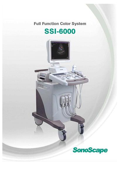
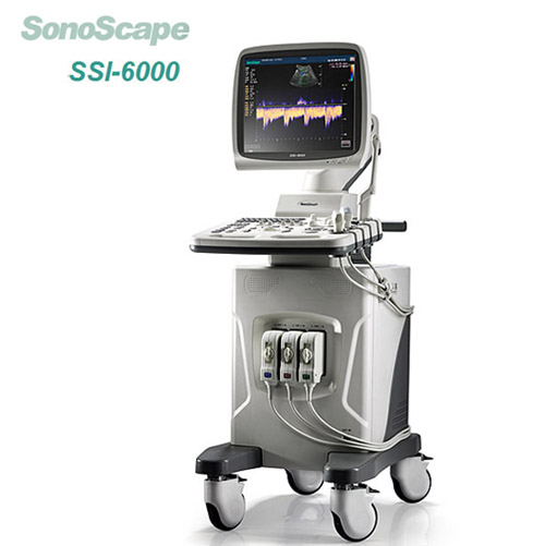
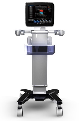
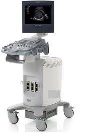
Thông số kỹ thuật chính
Advanced Technologies
Digital Front-End
Dual-beam Processing
Compound Imaging
Micro Scan Processing
Harmonic Imaging
High Pulse Repetition Frequency
Panoramic imaging
4D imaging
Graphic Exam Icon
Standard components
Color Mode
PW Mode
CW Mode
THI Mode
3D Mode
Dicom
Cardic Package
OB Package
Urology Package
Vascular Package
Small Part Package
MLA Probe
Tee Probe
Phased Array Probe
12MHZ Probe
ECG Support
DVD Support
Steer M Support
TDI support
HPRF support
Dual Beam support
u-SCAN support
Panoramic support
Color M Support
Optional Functions
4D support
MO Drive Support
B Flow Support
Dicom Wklist Support
IMT Support
System Overview
Applications
Abdominal
CardiologySpecifications for SSI-6000 High-Performance Color System
9034-0081-01D
Page 6 of 19
Obstetrical
Gynecological
Musculoskeletal
Vascular
Urological
Small Parts and Superficial
Pediatric
Anaesthesia
Scanning Methods
Electronic Convex Sector
Electronic Linear
Electronic Phased Array Sector
Sweep Angle
Curved Probe:70 degree or more
Phased Array Probe:90 degree or more
Micro-curved Probe:193 degree or more
Transducer Types
Convex Array
Micro convex Array
Linear Array
Phase Array
Operating Modes
B-Mode
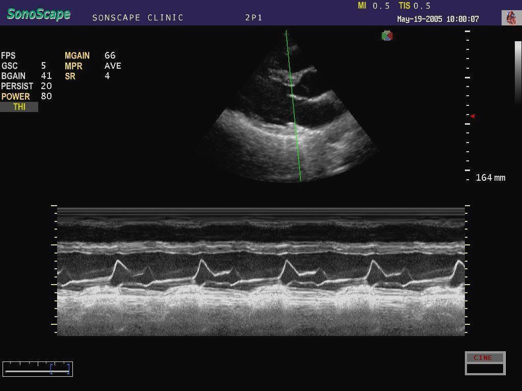
M-Mode
TDI-Mode
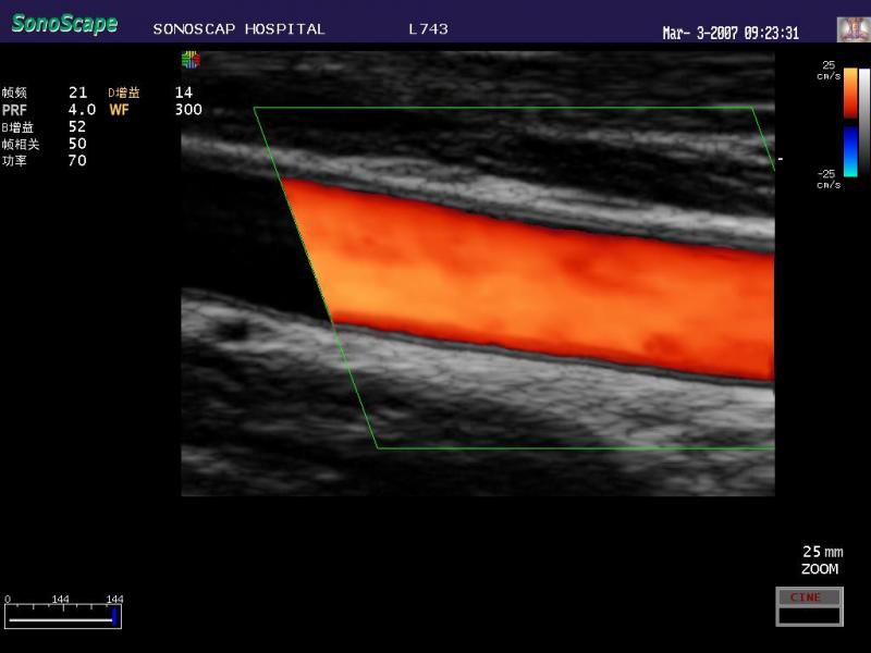
Color Flow Mode(CFM)
Power Doppler Imaging(PDI)
Pulse Wave Doppler(PWD)
Continuous Wave Doppler(CWD)
3D/4D Mode
Color M Mode
Steer M-Mode
Display Modes
B: Gray-scale imaging
Color: Color Doppler, Power Flow and
Directional Power Flow Imaging
THI(Tissue Harmonic Image)
Dual B, Quad Display
B and M, display format selectable
B and Doppler
B+Color
Dual B(Flow)
Triplex mode: B, Flow, and PW/CW
Doppler
B, Flow, and Color M
Simultaneous Refresh Display
Variable screen size: Change the screen
ratio of 2D and Doppler/M in duplex or
triplex mode
Panoramic Imaging
Compound Imaging
Trapezozd Imaging
Standard Features
Frame Rate:Max.510 frames/sec or moreSpecifications for SSI-6000 High-Performance Color System
9034-0081-01D
Page 7 of 19
Display Gray Scale:256 levels
Digital Channel Number:1024
Probe Elements: Up to 256
Media &Peripherals
Color Desk Jet Pinter(optional)
HP5850/6840/6848/6940/K5400dn
B/W Video Pinter(optional)
UP895MD
Color Video Pinter(optional)
Sony UP-20
System Menu Setting
File Manager
Copy、Paste、Delete
Convert to PC Format
Display Report Files Only
Display Image Files Only
Multiple Selection
Search
Set Time/Date
Facility Name
Dicom
System Information
Control Number
Software Version
System Setting
General Setting
Language Setting
English
Simple Chinese
Spanish
Russian
French
Italian
Screen Saver
Trackball Sensitive
Clip Format
CIN
WMV
AVI
Date Format
mm/dd/yyyy
yyyy/mm/dd
dd/mm/yyyy
Caps Lock:on/off
Print Size
Still Format
PPM
JPG
BMP
TIF
Set Printer
Printer Driver
Video Invert
Insert Driver
Set Calculation Menu
2D Mode
Angle
VolumeSpecifications for SSI-6000 High-Performance Color System
9034-0081-01D
Page 8 of 19
Volume L×W×H
Doppler Area
Vascular
Small Part
Obstetrical/ Gynecological
Left Ventricle
Urologic
Mitral Valve Diam
Lv Outflow Diam
Pul.Valve Diam
PW Mode
Flow Velocity
Acceleration
Time
Heart Rate
Cardiac
Obstetrical/ Gynecological
Vascular
M Mode
Distance
Time
Slope
Heart Rate
left Ventricle
Mitral Valve
Aortic Valve
Set Measurement Method
BPD Method
Hadlock
Jeanty
FL Method
Hadlock
Hohler
Jeanty
CRL Method
Robinson
Hadlock
Nelson
EFW Method
WEI/SAB HC,AC,FL
Shepard AC,BPD
Hadlock1 AC,FL
Hansman AC,FL,HC
Tokyo BPD,APTD,TTD,FL
Hadlock2 HC,AC,FL
Hadlock3 BPD,AC,FL
Hadlock4 HC,AC
Hadlock5 BPD,HC,AC,FL
Shinozuka BPD,AC,FL
Warsof FL,AC
BSA setting
Eastern
Western
Measure Method
Ellipse
Trace
Package
All Package
Icon Driven
Continue Dist:on/offSpecifications for SSI-6000 High-Performance Color System
9034-0081-01D
Page 9 of 19
Dop Auto
AUTO
SEMI-AUTO
Define OB table
Create
Edit
Delete
Annotation Edit
Insert
Delete
Edit
Save
Define quick key
GS (Gestational Sac diameter)
CRL (Crown Rump Length)
BPD (Biparietal Diameter)
HC (Head Circumference)
AC (Abdominal Circumference)
FL (Femur Length)
NT (Nuchal Translucency)
Load Default
Load
Create
Retrieve
Post-Processing
RAW data digital processing
Read Zoom up to 10x
B Mode
GSC
Chroma
LT RT
Play/Stop
Loop Speed
Start
End
Frame By Frame
Color Flow Mode
C Map
B Reject
Flow Invert
Loop Speed
Start
End
Frame By Frame
PW/CW -Mode
Chroma
Video Invert
Display Format
Start
End
Frame By Frame
Baseline
M-Mode
Chroma
Video Invert
Display Format
Start
End
Frame By FrameSpecifications for SSI-6000 High-Performance Color System
9034-0081-01D
Page 10 of 19
Scanning Parameters
B-Mode
Focus: Up to 12, focus span adjustable
zoom: Max.>= 10,Show zoom X value
TGC(Time Gain Control) 8 slide controls
Tissue acqustic: Adjustable according to
tissue type (1400-1700,10steps each)
Dynamic range-compression selections:
20-280 (probe dependent)
Gain:0-255 adjustable
Depth: 32.9 cm Max (probe dependent)
GSC(gray scale curve) 7 steps selectable
Persist (Frame correlation): 0-95 (probe
dependent)
Chroma: Max.13 selectable
SEC.WIDTH: B Image width adjustable
SEC.POS: B image lateral position
adjustable
Line Density: 3 selections (high/med/low)
Adapt . IM Fusion: 15 kinds
Biopsy Guide: on/off
Biopsy Offset adjustable
Biopsy Angle adjustable
Left and Right Inversion
Up and Down Inversion
Trapezoid Image: ON/OFF(liner array
probe)
u-Scan: adjustable
Compound Image: ON/OFF
Frequency : 5 steps
Power: 1 to 100 changeable,1 steps each
B Steer Mode
Color Flow Mode/TDI Mode
Triple bean functin
Frame Rate: max 25 frames/sec
Color Area Size and Position: adjustable
Persistence: 0-80(probe dependent)
Frequency Range: 5 steps
Pulse Repetition Frequency: 0.5-12KHZ
Steer Angle: 5 kinds (linear probe )
Max. ±20 degrees,
0, ±16, ±20 changeable
Baseline: ±15 steps
Filter: Up to 750 Hz (exam dependent)
B and B(Flow) Simultaneous Real-time
Display
Color Map: 6 kinds
Imaging Area and Position (adjustable)
B Reject: 0-255
Flow Invert: ON/OFF
Left /right: ON/OFF
Line Density: 2 kinds (low and high)
Color Distribution Display in Freeze Mode.
M-Mode
sweep speed: 2、4、6、8sec/plane
Chroma: 5 kinds selectable
Video Invert: ON/OFF
Frequency: 5 steps
M Process: Switch average or peak Specifications for SSI-6000 High-Performance Color System
9034-0081-01D
Page 11 of 19
detection processing for the M vector
display.
Steer M: 3 lines,Display Frame Rate
Display format: H1/2、H1/4、V1/3、V1/2、
V2/3、O1/4
Spectral Doppler
Doppler methods
PW (pulsed wave) Doppler
CW Doppler
One Button Optimization function
Doppler Envelope function in Real time
High Pulse Repetition Frequency
PW: 1-20KHz (exam dependent)
CW: 1-24KHz
Max velocity range:
0.0004-40.9 m/s(pw)
0.0013-49.1 m/s(cw)
BaseLine Shift: Available up to 17 steps
Angle correction: 0-80 degrees
Dynamic Rage: 10 steps selectable
Steer Angle: 5 kinds (linear probe )
Max. ±20 degrees,
0, ±16, ±20 changeable
Spectrum Inversion: Possible
Angle Correction: on /off
Sample Volume Size for PW Doppler:
1 -20 mm, changeable in 1 mm step
Sweep Speed: 2、4、6、8sec/plane
Chroma: Max.5 Kinds adjustable
Video Invert: on/off
2D Refresh: on/off
Display format: H1/2、H1/4、V1/3、V1/2、
V2/3、O1/4
3D/4D Mode
3 arbitrary sections simultaneously
Clear Roi
Restore Roi
Crop: on/off
Roi Mode: on/off
Hide Roi: on/off
Render Mode: Vol、MaxIP、X-ray
Auto Rotate (45、90、180、270、360
degrees adjustable )
Trace Cut: on/off
Undo Cut
Clip Plane: on/off
Opacity Offset: 0-255 adjustable
Opacity Slope: 0-255 adjustable
Multi-slice:Ref A、Ref B、Ref B
Slice Spacing:0.5-2.0 adjustable
Scan Method: Lin、Sec
Z Scale: adjustable
Z Angle: 10-170°adjustable
Zoom
Display mode
Dual Display
Quad Display
Full Display 3DSpecifications for SSI-6000 High-Performance Color System
9034-0081-01D
Page 12 of 19
Full Display 4D
Cine Review: on/off
Sweep Angle:20-75 degrees
Rescan: on/off
Image Quality: high、med、low
4D Gain: adjustable
Frame Rate: 5 frames/sec or more
Print
full scan of the Region of interest
Save images
Integrated Data Management
System
Hard Disk memory capacity: 160 G,
Storage media
USB Drive
DVD Drive
Storage of Images and Cine
Cine loop: 10000 frames or more
Cine loop time:60 seconds or more
Real time single/dual static and dynamic
Image storage
Archived image can be viewed on PC
Cropboard function: in Freeze Mode
Cine play back mode for Dop.
Doppler Cine Sound Play Back Function
DICOM Network Communication
Conformity to DICOM Standard: Service
class user of storage, (for details, please
refer to the DICOM conformance
statement issued by SonoScape.)
Storage: Directly transmits image with
patient information to a DICOM file server
Physiological Signal Display
ECG, Pulse wave
ECG Lead-three lead system
ECG Gain: adjustable
ECG Position: adjustable
ECG Invert:on/off
R-Trigger:on/off
Trigger Delay: adjustable
Frame Count: adjustable
User Interface
Operator Keyboard
Alphanumeric Keyboard
Shortcuts Keyboard
Integrated Recording Keys for Remote
Control of Peripheral Devices and DICOM
Devices
8 TGC Pods
Integrated function key
Character and icon
Character Input Area: ID, Name, DOB,Sex,
Weight, Height, LMP etc
Body Mark:52 kindsSpecifications for SSI-6000 High-Performance Color System
9034-0081-01D
Page 13 of 19
Electrical Power
Voltage:100/220 Volts AC
Current: 3.15 Amps
Frequency:50/60Hz
Dimensions and Weigh
502mm(W)×807mm(D)×1326mm(H)
Approx.64.5kg (main unit only)
Display Screen
17-inch High-Resolution Color LCD
monitor
Contrast and bright: 0-100 changeable
Environmental Requirements
In operation
Temperature:+10 to +40 degrees C
Relative Humidity: 30% to 75%
(non condensing)
Atmospheric pressure: 700 to 1060hPa
In Storage/Transportation
Temperature: -20 to +55 degrees C
Relative humidity: 20%- 90%
(non condensing)
Atmospheric Pressure: 700 to 1060hPa
Probe Connectors
Active Connectors: 3 connectors
Optional Probe
Phased Array Probe(Cardiology)
2P1 (1.9-6 MHZ)
5P1 (4.2-11 MHZ)
Linear Probe(Vascular, Small Part)
L741 (5-16 MHZ)
L742 (4.5-15 MHZ)
L743 (5-16 MHZ)
L752 (4.5-15 MHZ)
10L1 (4.5-15 MHZ)
L541 (3.7-8 MHZ)
Curved Probe(Abdomen, OB/GYN)
C344 (2-7 MHZ)
C362 (2-7 MHZ)
C542 (3.7-11 MHZ)
Micro-curved Probe(Transvaginal)
6V1 (3.9-15 MHZ)
6V3 (3.9-15 MHZ)
Micro-curved Probe(Cardiology)
C311 (2-6 MHZ)
C611 (4-13 MHZ)
4D Probe
VC6-2(2-7 MHZ)
Linear,Surgical(Surgery)
10I2 (4.5-15 MHZ)
Measurements/Calculations
General Measurements/Calculations
On B-Mode
Distance(real time、freeze)
Area and circumference (Trace, Ellipse)Specifications for SSI-6000 High-Performance Color System
9034-0081-01D
Page 14 of 19
(real time、freeze)
Volume (L×W×H,Area×L)
Angle
On M-Mode
Velocity
Distance
Time
Heart rate
Slope
On Spectral Doppler
Time Interval
Velocity
Velocity Ratio
Velocity Time Integral
Heart Rate
Velocity
Acceleration
Resistance Index
Pulsatility Index
Pressure half time
PV(peak Velocity)
Mean Flow Velocity
End diastolic Velocity
PG((Pressure gradient)
Auto Trace
Manual trace
On Color Mode
Color Flow Velocity
Doppler Area
proximal Isovelocity surface area
On 4D-Mode
Distance
Area and circumference
Volume
Obstetrical/ Gynecological
Measurements & Calculations
B Mode
GS (Gestational Sac diameter)
CRL (Crown Rump Length)
BPD (Biparietal Diameter)
HC (Head Circumference)
AC (Abdominal Circumference)
FL (Femur Length)
CER (Cerebellum)
OFD (Occipitofrontal Diameter)
Fibula (Fibula Length)
Foot (Foot Length)
AA (Abdominal Area)
APAD (Anteroposterior Abdominal
Diameter)
HA (Head Area)
Humerus (Humerus Length)
Kidney (Kidney Length)
APTD (Anteroposterior Trunk Diameter)
OOD (Outer Orbital Diameter)
Radius (Radius Length)
TAD (Transverse Abdominal Diameter)
TC (Thoracic Circumference)
THD (Thoracic Diameter)Specifications for SSI-6000 High-Performance Color System
9034-0081-01D
Page 15 of 19
Tibia (Tibia Length)
TTD (Transverse Trunk Diameter)
Ulna (Ulna Length)
Umb VD (Umbilical Vein Diameter)
NT (Nuchal Translucency)
LV (Lateral Ventricle)
UT L (Uterus Length)
UT H (Uterus Height)
UT W (Uterus Width)
Cx (Cervix)
En-T (Endometriosis)
Rt OV L (Right Ovary Length)
Rt OV H (Right Ovary Height)
Rt OV W (Right Ovary Width)
Lt OV L (Left Ovary Length)
Lt OV H (Left Ovary Height)
Lt OV W (Left Ovary Width)
AFI (Amniotic Fluid Index)
HIP(Hip Joint)
Dominant Follicle
EFA(Estimated Fetal Age)
EDD(Estimated Date of Delivery)
EFW ( Estimated Fetal Weight)
AUA(Average Ultrasound Age)
Fetal HR(Fetal Heart Rate)
PW Mode
Umb A (Umbilical Artery)
MCA (Middle Cerebral Artery)
Rt Uterin A (Right Uterine Artery)
Lt Uterin A (Left Uterine Artery)
Fetal AO (Fetal Aorta)
Cardiac measurements
B-Mode
Left Ventricular Fuction Measurement
Single Plane Ellipse Method
LVALd: Left Ventricular Long-axis
Area at end Diastole
LVLd: Left Ventricular Long-axis
Length at end Diastole
LVALs: Left Ventricular Long-axis
Area at end Systole
LVLs: Left Ventricular Long-axis
Length at end Systole
Biplane Ellipse Method
LVALd: Left Ventricular Long-axis
Area at end Diastole
LVALs: Left Ventricular Long-axis
Area at end Systole
LVAMd: Left ventricular short-axis
area at end diastole
LVIDd: Left ventricular short-axis
diameter at end diastole
LVAMs: Left ventricular short-axis
area at end systole
LVIDs: Left ventricular short-axis
diameter at end systole
Bullet
LVAMd: Left ventricular short-axis
area at end diastoleSpecifications for SSI-6000 High-Performance Color System
9034-0081-01D
Page 16 of 19
LVAMs: Left ventricular short-axis
area at end systole
LVLd: Left ventricular long-axis
length at end diastole
LVLs: Left ventricular long-axis
length at end systole
Simpson Method
LVAMD: Left ventricular short-axis
area at end diastole
LVAMs: Left ventricular short-axis
area at end systole
LVAPd: Left ventricular short-axis
area at the level of the
papillary muscle at end
diastole
LVAPs: Left ventricular short-axis
area at the level of the
papillary muscle at end
systole
LVLd: Left ventricularlong-axis
length at end diastole
LVLs: Left ventricular long-axis
length at end systole
Cube
IVSTd: Interventricular septal
thickness at end diastole
LVIDd: Left ventricular short-axis
diameter at end diastole
LVPWd: Left ventricular posterior
wall thickness at end
diastole
IVLTs: Interventricular septal
thickness at end systole
LVIDs: Left ventricular short-axis
diameter at end systole
LVPWs: Left ventricular posterior
wall thickness at end
systole
Teichholz
LVLDd: Left ventricular short-axis
diameter at end diastole
LVIDs: Left ventricular short-axis
diameter at end systole
Gibson
LVLDd: Left ventricular short-axis
diameter at end diastole
LVIDs: Left ventricular short-axis
diameter at end systole
Biplane Disk
Diastole 2CH
Diastole 4CH
Systole 2CH
Systole 2CH
Mitral Valve Diam
Lv Outflow Diam
Pul.Valve Diam
M-Mode
Left Ventricular Fuction Measurement
Cube
LVIDd: Left ventricular short-axis Specifications for SSI-6000 High-Performance Color System
9034-0081-01D
Page 17 of 19
diameter at end diastole
LVIDs: Left ventricular short-axis
diameter at end systole
LVPWd: Left ventricular posterior
wall thickness at end
diastole
LVPWs: Left ventricular posterior
wall thickness at end
systole
Gibson
LVLDd: Left ventricular short-axis
diameter at end diastole
LVIDs: Left ventricular short-axis
diameter at end systole
Teichholz
LVLDd: Left ventricular short-axis
diameter at end diastole
LVIDs: Left ventricular short-axis
diameter at end systole
Mitral Valve Measurement
Aortic Valve Measurement
PW-Mode
Mitral Valve Measurement
Aortic Valve Measurement
Tricuspid Valve Measurement
Pulmonary Valve Measurement
TEI Index Doppler Measurement
Vascular Measurements Calculations
ICA (Internal Carotid Artery)
ECA (External Carotid Artery)
CCA (Common Carotid Artery)
INT IL (Internal iliac)
EXT IL (External iliac)
ILIAC (Common iliac)
CFA (Common Femoral Artery)
PROFUN (Profunda)
LT CIR (Lateral Circumflex)
SFA(Superficial Femoral Artery)
POP (Popliteal Artery)
PTA (Posterior Tibial Artery)
PERON (Personal Artery)
ATA (Anterior Tibial Artery)
DR PED (Dorsalis Pedis)
%A REDUC (Area reduction percent)
%D REDUC (Diameter reduction
percent)
PI (Pulsatility Index)
RI (Resistive Index)
S/D (Systolic/Diastolic Ratio)
PG((Pressure gradient)
PV(peak Velocity)
IMT
Urological Measurements
&Calculations
Left Kidney
Right Kidney
Left-Renal Cortex
Right-Renal CortexSpecifications for SSI-6000 High-Performance Color System
9034-0081-01D
Page 18 of 19
Left-Adrenal Gland
Right- Adrenal Gland
Bladder Volume
Residual Urine
Urine Area
Urine Height
Whole Prostate Volume
Trans Zone Volume
Left-Seminal Vesicles
Right- Seminal Vesicles
Left-Testicle
Right- Testicle
Small Part Measurements
L-Thyroid
R-Thyroid
Thyroid Isthmus
L-Superior Parathyroid
L-Inferior Parathyroid
R-Superior Parathyroid
R-Inferior Parathyroid
Report functions
Obstetrical /Gynecological report
(revisability)
Obstetrical Curve
Fetal Anatomy
Biophysical Profile
Fetal Compare
Picture